
+86 18126862446
Products

PalmUS
PortableUS
TrolleyUS
VetUS
BladderUS
Ophthalmic
ECG,Monitor,etc.
Acc.
[pics:title]
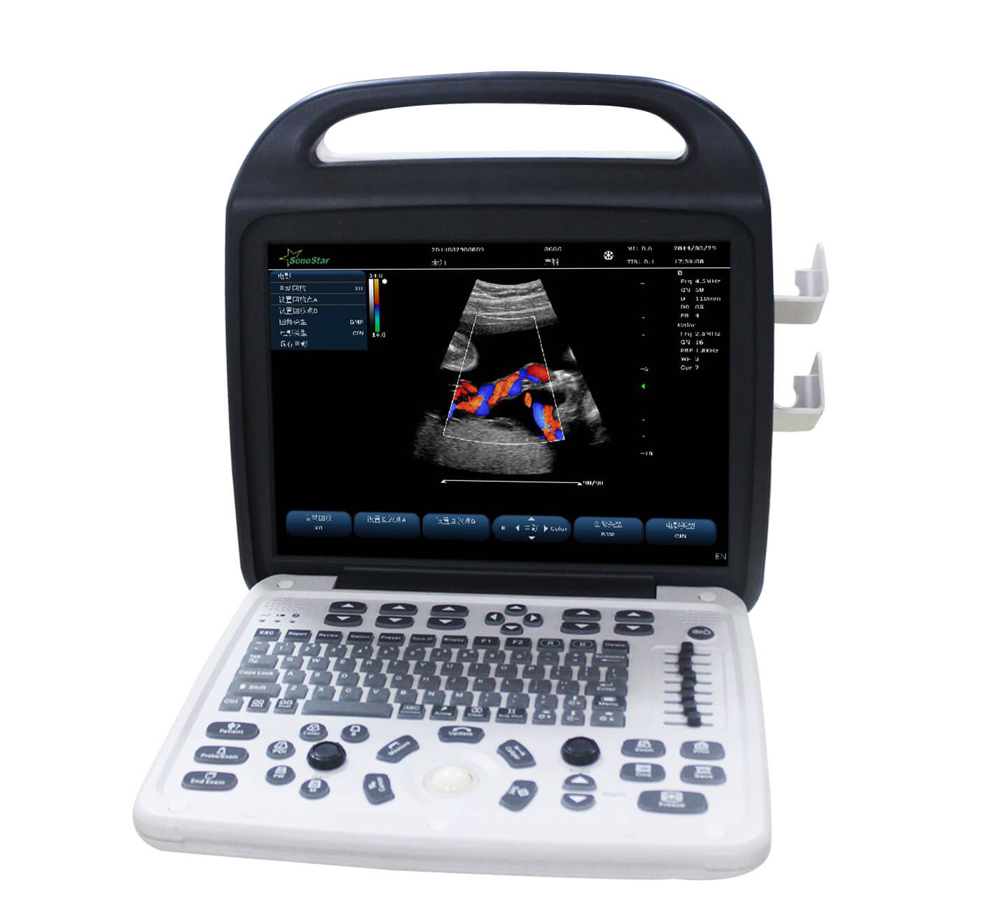
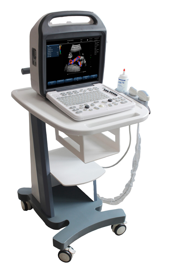
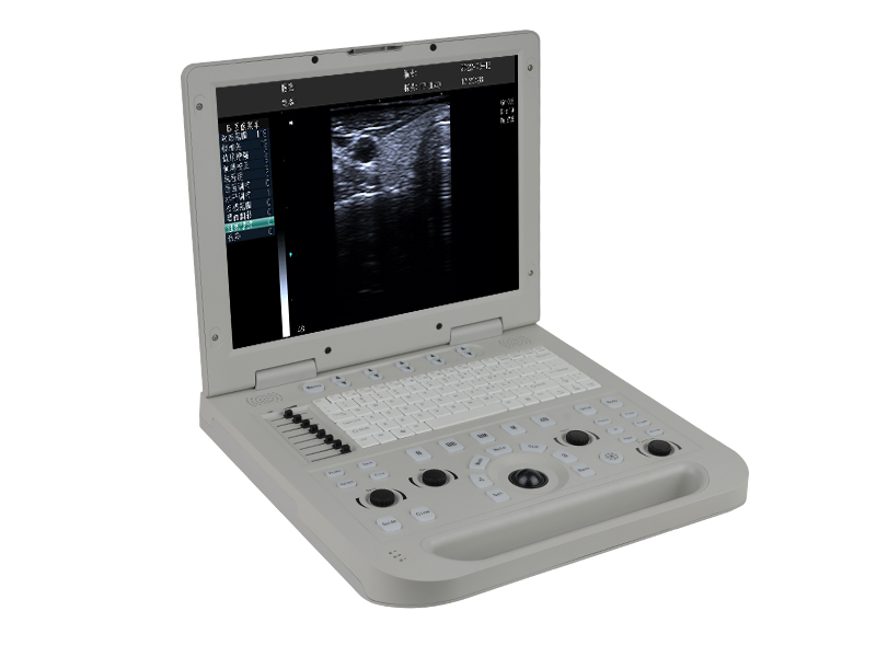
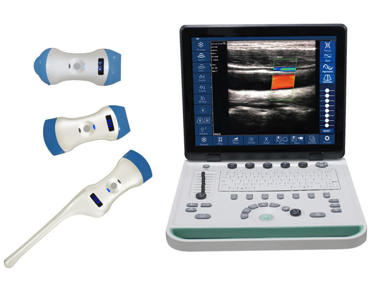
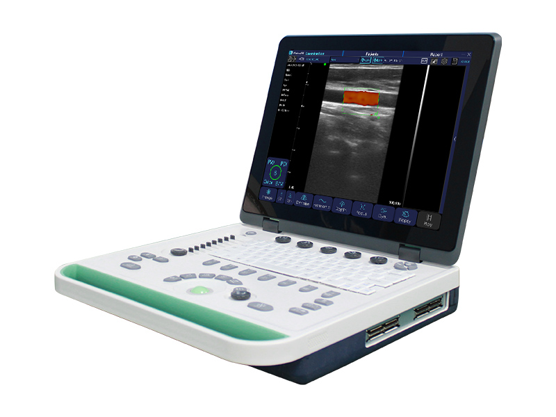
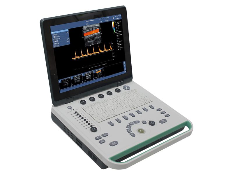
C10 Color Doppler Ultrasound Scanner
[pics:title]

C10 Color Doppler Ultrasound Scanner

15 inch screen, high cost-effective
Inquire →


1、Product Usage

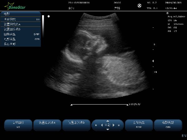
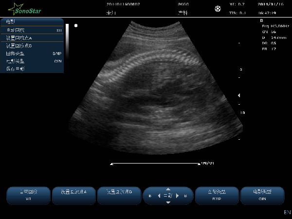
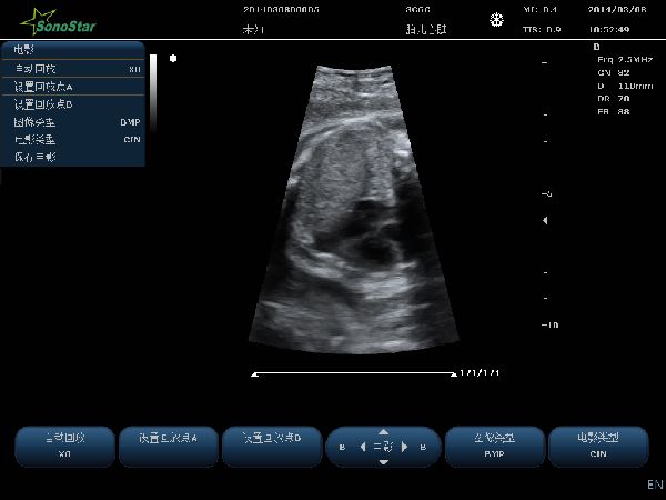
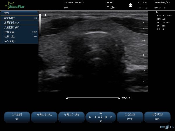
Suitable for systemic ultrasound applications including abdominal, gynecological, obstetric, cardiac, superficial tissue and small organs, neurological, peripheral vascular, cranial, urinary, pediatric, orthopedic, transrectal, and ultrasound-guided interventional procedures.


2、Host Specifications

2.1 ≥15-inch high-definition, medical-grade professional color LCD display. 

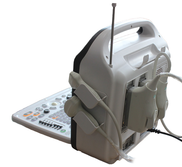
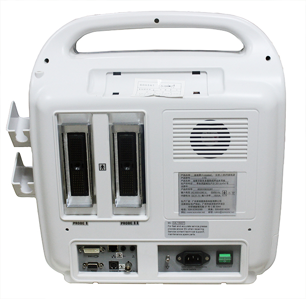
2.2Number of host probe ports: ≥2. Standard equipped with 2 probe ports.

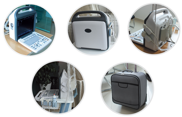
2.3 size and weight:

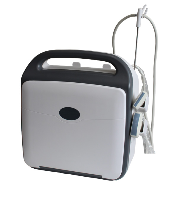
1. Machine size: 400mm(width) × 394mm(height) × 172mm(thickness)

2. Packaging size: 465mm × 465mm × 310mm

3. Machine weight: 7.5kg; 

4. Total package weight:  (machine + 2 probes + packing): 12kg

2.4 Battery parameters: Capacity ≥7000 mAh. After fully charged, the continuous normal operation time in scanning mode is ≥ 2 hours.

2.5 Safety Standards: Complies with commercial product safety and quality requirements.

2.6 Supported languages include English、Chinese (keyboard input, annotations, control panel, etc.).

2.7 Startup time:≤13 seconds. The main interface and images are displayed within 13seconds.

2.8 Supports system upgrades from the bootloader.

2.9 Built-in 12-lead electrocardiogram ECG machine(Optional)


3、Imaging Control

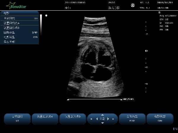
3.1 Imaging Modes:B、M、PW、Color、PDI;

3.2 Display Modes:B、dual、quad、B+M、M、B+Color、B+PDI、B+PW、PW、B+Color+PW、B+PDI+PW、B/BC real time 

3.3 Digital Imaging Technologies:Multi-beam formation、Real-time point-by-point dynamic focusing imaging、Pulse inversion harmonic imaging、Spatial compound imaging、Image enhancement、adjustable at multiple levels

3.4 Real-time scanning for images (B, B+C, 2B, 4B), supporting stepless zoom.

3.5 Automatic image optimization, applied to 2D, color, and spectrum modes.

3.6 Spectral Doppler Display and Analysis System

3.7 Puncture Guidance Function: B-mode image supports the display of a puncture line,  and supports the display of a 0-angle puncture guidance line.

3.8 B-mode image supports the display of a lithotripsy targeting line and a PICC guidance line.


4、Measurement and Analysis: (B-mode, M-mode, Spectral Doppler, Color Doppler)

4.1 General Measurement (Distance Measurement, Ellipse and Trace Measurement for Area and Perimeter, Volume Measurement)

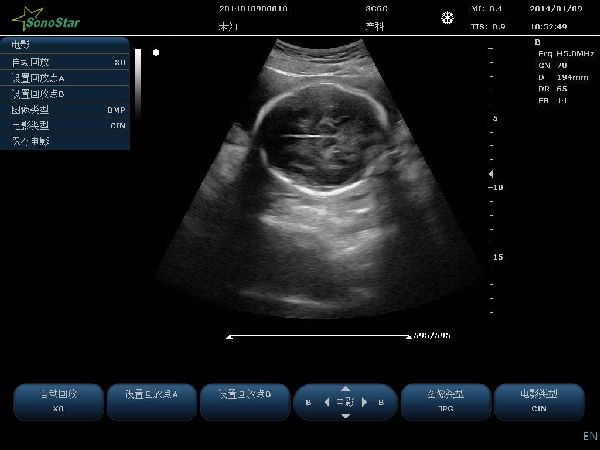
4.2 Obstetric and Gynecologic Measurement: Dedicated obstetric/gynecologic measurement and analysis, including fetal biophysical profile (FBP) and obstetric formulas for the Chinese population.

4.3 Cardiac Function Measurement

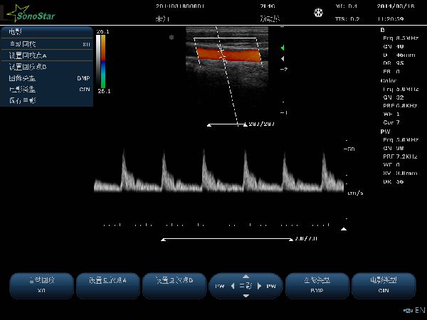
4.4 Peripheral Vascular Blood Flow Measurement and Analysis Report Function

4.5Optionally equipped with automatic Intima-Media Thickness (IMT) measurement.

4.6Doppler Measurement and Analysis (automatic and manual trace measurements with automated calculation of parameters).


5、Probe Specifications

5.1 Frequency: Wideband frequency probe, B/M and C/D independent frequency conversion

5.2 Convex array, linear array and phased array probes all have ≥5 frequency conversions

5.3 Puncture guidance: Puncture guidance device is optionally available for all probe types.


6、Imaging Parameters

Preset Conditions: Pre-optimized imaging presets are configured for specific examination organs.

6.1 2D Grayscale Imaging Mode

6.1.1 Grayscale Mapping: ≥15 levels

6.1.2 Frame Correlation: ≥8 levels

6.1.3 Image Enhancement: ≥5 levels

6.1.4 Spatial Compounding: Adjustable (On/Off)

6.1.5 Scan Density: High, Medium, Low

6.1.6 Image Flip: Up/Down, Left/Right

6.1.7 Maximum Display Depth: ≥350 mm

6.1.8 Maximum Number of Transmit Foci: ≥4

6.1.9 Maximum Number of Transmit Focus Positions: ≥16

6.1.10 B-mode Maximum Frame Rate: ≥400 frames/sec

6.1.11 TGC: ≥8 segments

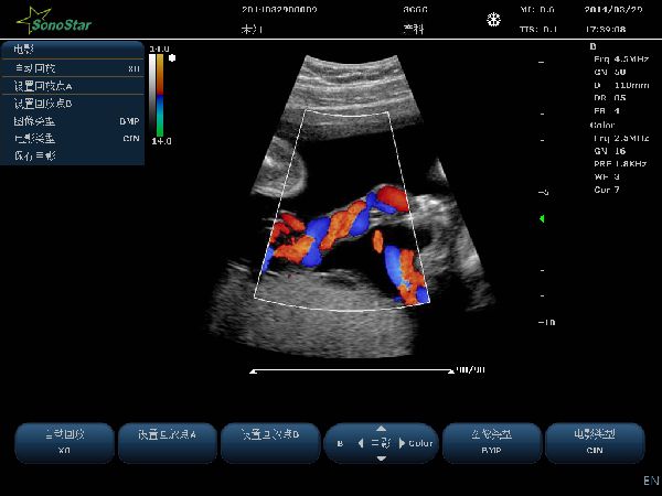
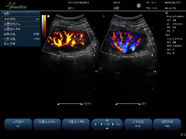
6.2 Color Doppler Imaging Parameters

6.2.1 Color Steering: ≥±12 degrees (for linear transducers)

6.2.2 Doppler Frequency: ≥2 independently adjustable and visually selectable ranges

6.2.3 B/Color Dual-Screen Real-Time Display

6.2.4 PRF: ≥15 steps, 0.6 kHz to 11.7 kHz

6.2.5Color Map: ≥4 types

6.2.6 Color Correlation: ≥8 steps

6.2.7 Post-processing: ≥4 steps

6.3 Spectral Doppler Mode

6.3.1 Size/Position: Sample Volume (SV) size adjustable from 1.0 mm to 8.0 mm

6.3.2 PRF: ≥16 steps, adjustable from 0.7 kHz to 9.3 kHz

6.3.3 Scanning Speed (Sweep Speed): ≥5 adjustable levels

6.3.4 Correction Angle: -85° to 85° with 5° step size

6.3.5 Spectral Inversion: On/Off adjustable

6.3.6 Wall Filter: Adjustable with 4 or more settings

6.3.7 Doppler Audio: ≥20 levels

6.3.8 Supports automatic spectrum measurement.

6.3.9Supports blood flow measurement

6.3.10 Steering Angle: ≥ ±12° (for linear probe)

6.4 M mode

6.4.1 Sweep Sleep:≥ 5 levels

6.4.2 Line Average:≥8 levels


7、Cine Review

7.1 Available in all modes.

7.2 Supports manual and automatic playback (cine loop), with adjustable playback speed for automatic mode.

7.3 Max. Cine Storage Capacity:

B-mode: ≥5000 frames; Color/PDI Max.: ≥2300 frames

M/PW Max.: ≥190 s

7.4 Image Save:

single-frame formats: BMP, JPG, FRM

Cine files include: AVI, CIN

(Supports offline analysis: After reloading stored or exported image data, it supports playback, measurement, parameter adjustment, annotation addition, and other operations.)


8、Built-in image and report management system

8.1 Digital ultrasound image storage capacity ≥128 GB

8.2 Integrated workstation enables synchronized preview of patient records and corresponding images.

8.3 Supports one-touch operation for direct image saving to the hard drive.

8.4Supports FTP transmission: Connect the ultrasound machine to a computer via an Ethernet cable to send exam data to an FTP server.

8.5 Supports USB export and import: Enables transferring exam data and images to a USB drive, as well as importing exam data and images from a USB drive.

8.6 Supports loop playback of selected exam images.

8.7 Supports report editing, report printing, and  report templates.


9、Input/Output

9.1Video Output:Composite Video, RGB, HDMI

9.2 Supports network storage: Transfer device data to a PC via Ethernet cable.

9.3Supports DICOM 3.0 connectivity: Automatically uploads patient data to the DICOM server upon exam completion.

9.4 Supports standard USB foot switches;

9.5 Supports USB keyboard and mouse;

9.6 USB Barcode Scanner Support;

9.7 Report Printer: Supports both monochrome/color laser printers and color inkjet printers; at least 3 models available for each type.

9.8 Video Printer: Supports monochrome and color video printers,supports digital video printers.


10、Optional probes

Probe name Probe type frequency range

3C6C      Convex array                2.5-6.0MHz

7L4C      Linear array                  6.5-10 MHz

10L25C   Linear array                  9.0-14.5MHz 

6E1C     transvaginal Convex array   5.0-9.0 MHz

6C15C    micro convex array          5.0-9.0MHz

6C11C    micro convex array          5.0-9.0MHz

7I4C      transrectal Linear array      6.5-10 MHz

2P2F     Phased array                 2.0~3.5MHz

5P2F     Phased array                 4.0~6.5MHz


Contact

Your Name :*

Tel Number:*

E-mail:

Company Name

Description

ADD:Guangzhou, China.  TEL:+86-20-32382095  PHONE:+86 18126862446  EMAIL:sonostar@sonostar.net  WEBSITE:https://www.sonostarmed.com
    XML MAP  HZSZNET.COM